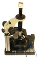
The CombiScope Scanning Probe Microscope (SPM) is an advanced research instrument that provides the entry path for researchers in materials science, biology, spectroscopy and photonics. If you work with transparent samples either in air or in liquid towards nanoscale structures and near-field optical properties investigation, the CombiScope is the right solution for you. It perfectly combines inverted optical and atomic force microscopies and unleash all the power of both techniques providing the instrument adjustment and measurement automation, high resolution and integration flexibility only available from AIST-NT.
Extraordinary productivity and easy operation
CombiScope is equipped with the fully motorized cantilever holder and photodiode positioning that provides the automated click-on-a-button laser-to-tip alignment. This option dramatically simplifies the entire system adjustment process and provides the highest level of system adjustment reproducibility. In addition, after you installed a new cantilever of the same or even different type, the same spot (within a few microns repeatability) on your sample surface can be easily found and scanned without any extra searching steps.
Top level scanner
The CombiScope utilizes the closed loop, high-dynamics, 3-axis piezo-nanopositioning scanner from the leader in precision motion control, Physik Instrumente. The top level scanner is the heart of the system which enables it to achieve very high levels of linearity, highest possible stiffness and extremely high precision motion.
IR AFM laser
The use of 1300 nm AFM laser eliminates any interference with VIS light-sensitive biological and semiconductor samples. It also makes it possible to perform simultaneous AFM and fluorescence or Raman scattering measurements without any crosstalk for most popular UV-VIS-NIR (364-830 nm) excitation lasers.
Integration with optics
Besides integrated inverted optical microscope such as Nikon Eclipse Ti-U and Olympus IX-71 with Phase Contrast and DIC, the CombiScope can be equipped with the head which provides the top and side simultaneous optical access with planapochromat objectives (100x, NA=0.7 and 10x, NA=0.28 respectively). This option opens up the way to combine the upright and transmission configurations to study transparent as well as non-transparent samples with optical, Raman and scanning probe microscopy techniques.
Solutions to work in liquid
The standard CombiScope's sample holders accommodates all common sample substrates, including slides, cover slips and 35mm Petri dishes. The specially design liquid cell with heating and liquid perfusion capabilities enables for biological samples to be delicately maintained in their physiological environment and at temperatures up to 60°C.
Top, side and bottom optical access
to the tip-sample area is provided to be able to explore the full capabilities of TERS using IR, VIS and UV high NA planapochromat objectives (top objective: up to 0.7 NA; side objective: up to 0.55 NA), which enable confocal detection of optical signal from the sample surface in a wide spectral range and the minimum size of excitation laser spot area. The properly designed side optical channel plays an extremely important role in successful TERS experimentation as it provides a much more significant Z component of the optical field and effectively excites the Plasmon resonance in the tip-sample junction.
All operating modes in one single instrument
The CombiScope comes with all modern AFM operating modes in one single instrument, without any extra costs and units, including such application-specific modes as force and electric nanolithographies, piezoelectric force microscopy (PFM), Kelvin Probe Microscopy and frequency modulation AFM (dynamic force microscopy with built-in PLL). In addition, the scanning tunneling microscopy (STM) head and Conductive AFM unit operating in the range 100 fA ÷ 10 uA (with 1 nA, 100 na and 10 uA subranges software switchable and current noise of 60 fA RMS for 1 nA subrange) and near-field optical microscopy (SNOM) head are available as the options. Such exceptional versatility of the instrument makes it a perfect solution for nanoscience.